|
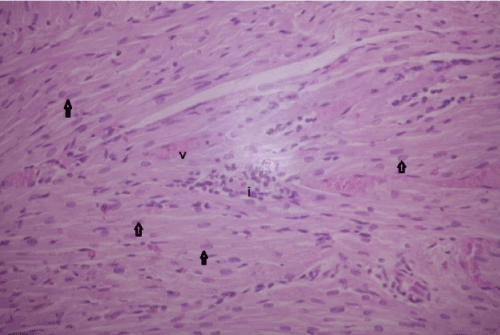
| Figure 7: A Higher magnification of a section in the cardiac muscle from treated group (group III) showing most of cardiac myocytes appearing normal with central oval nuclei (arrows) and presence of extravasation of blood between muscle fibers (v). Notice presence of mononuclearcellular infiltration (i) (H&E 200X). |