|
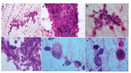
| Figure 2: (a) Chordoma smears at low power observed a few cells in a myxoid background (H&E 200X). (b) Moderate cellularity observed with predefined of mixed background (H&E 200X). (c) Singles cells demonstrates the seemingly syncytial appearance of this tumour cells (H&E 400X). And in (d), observed a high cellularity, cell with an epithelial appearance with abundant clear cytoplasm. The smear produces thick cytoplasmic bridges among a small cell group (H&E 400X). (e) Smear showed single cells with abundant clear and vacuolated cytoplasm and homogeneous nuclei (H&E 400X). (f) Close up observed two cell types of benign appearance (H&E 1000X). |