|
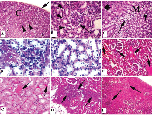
| Figure 3: A): Section of mature albino rats’ kidney of control group (I & II) showing normal, intact renal cortex (C), Bowman’s capsule (arrow head) and thin connective tissue capsule (arrow). H&E; (Obj.4X: Oc.10X). (B): Section of mature albino rats’ kidney of control group showing normal, intact renal corpuscle of glomerulus and bowman’s capsule (arrow), and are surrounded with proximal and distal convoluted tubules (arrow head). H&E (Obj.40X: Oc.10X). (C): Section of mature albino rats’ kidney of control group showing normal, intact medulla (M) of distal convoluted tubules (star), loop of henel (arrow) and connecting tubule (arrow head). H&E; (Obj.x 10: Oc.10X). (D): Section of mature albino rats’ kidney of exposed group to EMF (III) showing hyperemia with swelling in the linning epithelium of the glomeruli associated with a peri-glomerular focal area. H&E; (Obj.40 X: Oc.10X). (E): Section of mature albino rats’ kidney of exposed group to EMF (III) showing dramatic renal injury with tubular cell swelling with loss of brush border of convoluted tubules as well as necrosis. H&E; (Obj.40 X: Oc.10X). (F, G): Section of mature albino rats’ kidney of exposed group to EMF (III) showing dilatation and hyperemia in the intertubular cortical blood vessels (arrow). 6, 7) H&E (Obj.10X: Oc.10X). (H): Section of mature albino rats’ kidney of exposed group to EMF (III) showing leucocytic infiltration that are mainly of lymphocytes (arrow). H&E; Obj.10X: Oc.10X. (I): Section of mature albino rats’ kidney of group (IV) that kept for 48 days post the end of exposure to EMF without exposure showing the normal, intact renal corpuscles (arrow). H&E I) Obj.4X: Oc.10X.E; I) Obj.10X: Oc.10X; J) Obj.100X: Oc.10X; K) Obj.100X: Oc.10X. |