|
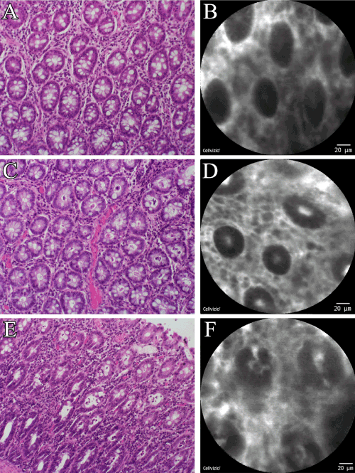
| Figure 4: Comparative images: standard histology versus confocal analysis. A blinded pathologist assigned an ischemia score to histological and confocal images. A) 20X Hematoxylin-eosin (HE), and B) confocal imaging score 0 (normal mucosa), C) 20X HE, and D) confocal imaging score 1 (submucosal congestion and swelling), E) 20X HE, and F) confocal imaging score 2 (diffuse swelling and necrosis of epithelium). |