 |
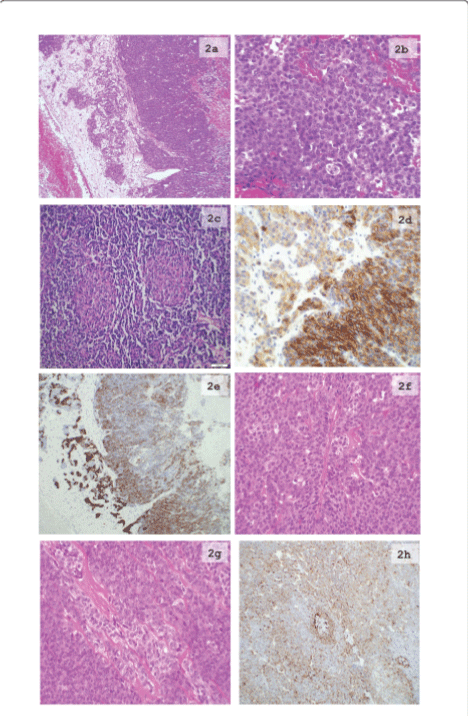
| Figure 2: Microscopic and immunohistochemical images from the primary tumor and recurrence. 2a: Low magnification of the tumor showing heterogeneous picture with different areas from the tumor with haemorrhages, small microcystic spaces and degenerative changes (H&E section magnification 10X). 2b: H&E section with solid proliferation of small atypical cells with round nuclei with “salt and pepper” chromatin and relative scarce eosinophylic cytoplasm (magnification 40X). 2c: H&E section demonstrating two squamoid bodies and partially proliferation of small cells with hyperchromatic nuclei and scant cytoplasm (magnification 20X). 2d: IHC staining with anti-CD56 antibody demonstrating strong membranous staining in the solid areas and weak focal, partial membranous staining in the acinar areas (20X). 2e: IHC staining with anti-Synaptophysin antibody demonstrating relatively weak staining in the solid areas and strong cytoplasmic staining in the acinar areas (10X). 2f: H&E section demonstrating solid proliferation and small microcystic spaces of small cells with hyperchromatic nuclei and scant cytoplasm (magnification 40X). 2g: H&E section showing different areas from the tumor with trabecular and microacinary growth pattern (magnification 40X). 2h: IHC staining with anti-Synaptophysin antibody demonstrating moderate diffuse staining in the solid areas (10X). |