|
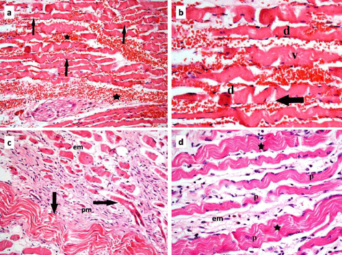
| Figure 2: Photomicrographs of skeletal muscle sections of rats in group II showing: In subgroup IIa: a] extravasated red blood corpuscles (*) among wavy darkly stained acidophilic and partially segmented muscle fibers (arrows); b] in higher magnification, multiple dark nuclei (d) and vacuolations (v) in the sarcoplasm of these fibers, with discontinuous myofibrils through most of its caliber (arrow). In subgroup IIb: c] some wavy darkly stained acidophilic muscle fibers (arrows). Few fibers have central nuclei. There is increased amount of endomysium (em) and perimysium (pm); d] multiple pale nuclei (p) in the darkly stained fibers. Many myofibrils are continuous but wavy (*) with increased amount of endomysium (em) (H&E, a and c 200X; b and d 400X). |