 |
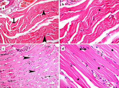
| Figure 3: Photomicrographs of skeletal muscle sections of rats in group III showing: In subgroup IIIa: a] a darkly stained acidophilic homogenous muscle fiber (arrow) and other normal slightly wavy fibers (arrowheads), and congested blood vessels (c); b] in higher magnification, fewer dark nuclei (d), and continuous but wavy myofibrils (*) compared to figure 3b. In subgroup IIIb: c] normal muscle fibers (arrowheads), infiltrating cells (arrows) among the fibers besides less congested blood vessels (v) compared to 4a. Note that many fibers show central nuclei; d] multiple pale nuclei (p) in normal muscle fibers, with continuous, straight and parallel myofibrils (*). An area of the sarcoplasm reveals transverse striations (arrow) and the vessels are less congested (c) (H&E, a and c 200X; b and d 400X). |