|
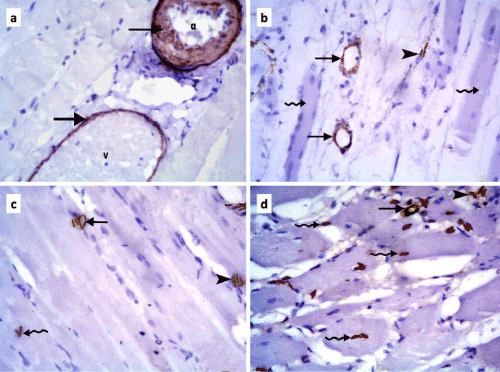
| Figure 4: Photomicrographs of skeletal muscle sections of rats in: a] control group showing +ve αSMA immunoexpression (arrows) in the wall of an arteriole (a) and a venule (v); b] subgroup IIb showing positive αSMA immunoexpression (arrows) in the wall of two small vessels and in the CT cells between (arrowhead) widely spaced muscle fibers (wavy arrows). Note the regenerating fiber with many large pale nuclei (left wavy arrow); c] subgroup IIIb showing +ve αSMA immunoexpression (arrow) in the wall of a small vessel and some +ve cells in the CT (arrowhead) between and among (wavy arrow) normal closely arranged muscle fibers compared to 5b; d] subgroup IIIb showing +ve αSMA immunoexpression (arrow) in the wall of a small vessel and multiple +ve cells in the CT between (arrowhead) and among (wavy arrow) disorganized muscle fibers. Note that some of the cells that overlapped muscle fibers gave +ve reaction while others did not (as compared to figure 4c). (αSMA immunostaining, 400X). |