|
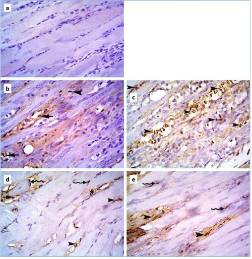
| Figure 5: Photomicrographs of skeletal muscle sections of rats in: a] control group showing negative CD34 immunoexpression among the fibers and the CT in between; b] subgroup IIb showing few +ve spindle cells inside blood vessels (arrow) and in the CT between (arrowhead) more or less regularly arranged fibers; c] subgroup IIb showing multiple +ve spindle cells in the CT between (arrowheads) widely spaced fibers; d] subgroup IIIb showing multiple +ve spindle cells inside a blood vessel (arrow), in the CT between (arrowheads) and among normal muscle fibers (wavy arrows) compared to 6b and 6c; e] subgroup IIIb showing numerous +ve spindle cells in the CT between (arrowheads) occasional widely spaced fibers and few +ve cells among the other fibers (wavy arrows) (CD34 immunostaining, 400X). |