|
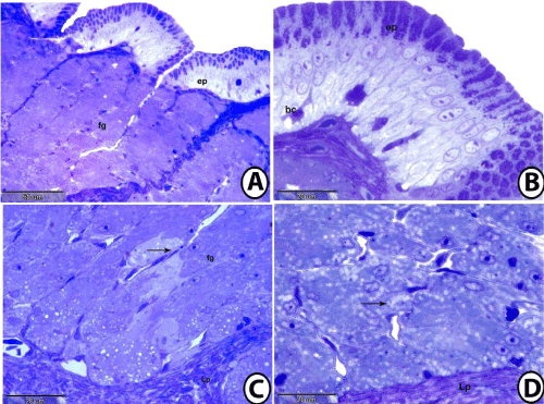
| Figure 4: semithin section of fundic mucosa stained with toluidine blue. 4A: Surface epithelium (ep) of fundic region lead to fundic glands (fg). Notice the abrupt transition from gastric pits to gastric glands. 4B: Surface simple columnar epithelium (ep) of fundic region showing positive reaction of the apical parts. Notice presence of undifferentiated basal cells (bc). 4C: lamina propria (Lp) contained fundic glands (fg) that opened into duct lined by simple squamous epithelium (arrow). 4D: lamina propria (Lp) of fundic region showing fundic glands that consisted of oxynticopeptic cells that contained many vesicles (arrow). |