 |
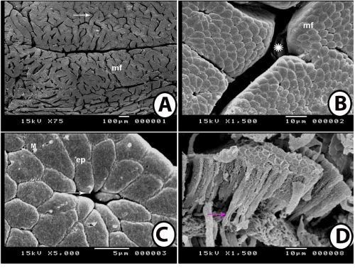
| Figure 5: scanning electron microscopy of the fundic region. 5A: primary mucosal folds (mf) of the fundic region of the stomach showing presence of large number of gastric pits (arrow). 5B: Scanning electron micrograph of the fundic region showing presence of large concavities (*) between the mucosal folds (mf). 5C: Scanning electron micrograph of the surface polyhedral shaped epithelial cells (ep) of the fundic region. Notice presence of gastric pits (arrow) and deposition of some mucous droplets (M) on the surface. 5D: Scanning electron micrograph of damaged surface of fundic region showing tall columnar surface epithelium with infranuclear region (arrow). |