 |
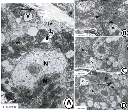
| Figure 6: Transmission electron microscopy on the gastric glands. 6A: Transmission electron micrograph of oxynticopeptic cell of the fundic glands contained nucleus (N), lysosomes (L), mitochondria (M), and secretory vesicles (V). Notice presence of short apical microvilli (arrow) and tubulovesicular network (tv). 6B: Transmission electron micrograph of the fundic glands showing 2 oxynticopeptic cells contained euchromatic electron lucent nucleus (N) with large nucleolus and large number of mitochondria (M) and secretory vesicles (V). 6C: Transmission electron micrograph of oxynticopeptic cell of the fundic glands contained nucleus (N), Golgi complexes (G), mitochondria (M) and secretory vesicles (V). 6D: Transmission electron micrograph of oxynticopeptic cell of the fundic glands contained nucleus (N), lysosomes (L), mitochondria (M), rough endoplasmic reticulum (rER) and secretory vesicles (V). |