 |
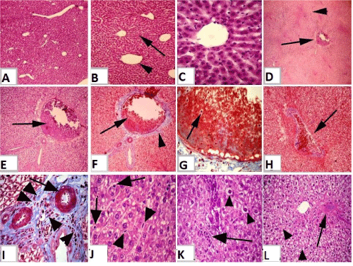
| Figure 1: (a): Section of mature Westar rats liver of control group (I) showing normal, homogenous, intact hepatic parenchyma; hepatic lobules, with normal central vein. H&E 4X:10X. (b): Section of mature Westar rats liver of control group showing normal hexagonal hepatic lobules with normal, regular radiated hepatic cords from the central vein to the peripheral of lobule (arrow), central vein (arrow head). H&E 10X:10X. (c): Section of mature Westar rats liver of control group showing normal hepatocytes of irregular polygonal shaped cells with single, central, large vesicular nucleus. Normal and regular hepatic cords that were dorsally radiating from the central vein. H&E 40X:10X. (d): Section of mature Westar rats liver of exposed group to tartrazine (II) showing severe steatosis, diffuse degeneration, necrosis of hepatic tissues with pale acidophilic hepatic parenchyma (arrow head) and with moderate disorganization of hepatic cords and sever congestion of portal vein (arrow). H&E 4X:10X. (e,f): Section of mature Westar rats liver of exposed group to tartrazine (II) showing sever congestion of portal vein (arrow) with fibrous tissue proliferation in the portal areas (arrow head). e) H&E, f) Blue Masson’s Trichrome, e) 10X:10X f) 10X:10X. g): Section of mature Westar rats liver of exposed group to tartrazine (II) showing sever congestion of portal vein that overfilled with erythrocytes and some lymphocytes (arrow). Blue Masson’s Trichrome 40X:10X. (h): Section of mature Westar rats liver of exposed group to tartrazine (II) showing sever congestion of portal vein (arrow) with fibrous tissue proliferation (arrow). Blue Masson’s Trichrome 10X:10X. i): Section of mature Westar rats liver of exposed group to tartrazine (II) showing dilated blood vessels of the portal area (arrow) with fibrous connective tissue proliferation and leukocytes infiltrations (arrow head). Blue Masson’s Trichrome 40X:10X. (j): Section of mature Westar rats liver of exposed group to tartrazine (II) showing the presence of Kupffer cells (arrow head) and lymphocytes (arrow) in between the necrotic hepatic cords. H&E 40X:10X. (k): Section of mature Westar rats liver of exposed group to tartrazine (II) showing enlarged necrotic hepatocytes with light and foamy cytoplasm that filled with vacuoles of variable size and mostly central pyknotic nuclei (arrow head) and leukocytes infiltrations (arrow). H&E 40X:10X. (l): Section of mature Westar rats liver of exposed group to tartrazine (II) showing wide area of necrotic hepatic parenchyma with light cytoplasm and mostly pyknotic nuclei (arrow head) and focal area of degenerated hepatocytes (arrow). H&E 20X:10X. |