 |
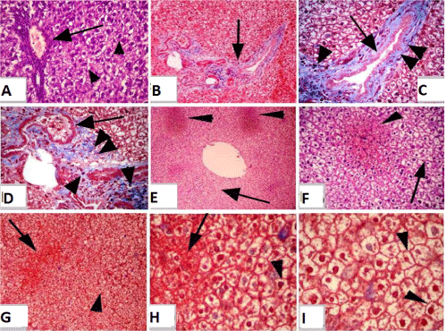
| Figure 3: (a,b): Section of mature Westar rats kidney of control group (I). (a): showing normal, intact renal tubules as well as renal glomeruli. (b): higher magnification of Figure 1 showing the same. (a,b) H&E a) 10X:10X b) 4X0: 10X. (c): Section of mature Westar rats kidney of exposed group to tartrazine (II) showing degenerative changes in the renal tubules (arrow head) and glomeruli (arrow). Blue Masson’s Trichrome Obj.x20: 10X. (d): Section of mature Westar rats kidney of exposed group to tartrazine (II) showing an enlargement of renal glomeruli with vacuolations (single arrow head) and separation of its lining and epithelial cells (double arrow head) as well as congestion of its blood capillaries (arrow). H&E 4X0: 10X. (e): Section of mature Westar rats kidney of exposed group to tartrazine (II) showing hypertrophy and degeneration of the lining epithelial cells of the renal tubules. H&E Obj.x20: Oc.x15. (f): Section of mature Westar rats kidney of exposed group to tartrazine (II) showing hyaline degenerative changes in the renal tubules (arrow head). H&E 4X0: 10X. (g): Section of mature Westar rats kidney of exposed group to tartrazine (II) showing fibrous connective tissue proliferation with leukocytes infiltration in between renal tubules (arrow) and hyaline degenerative changes in the renal tubules (arrow head). Blue Masson’s Trichrome Obj.x20: 10X. (h,i): Section of mature Westar rats kidney of exposed group to tartrazine (II) showing blood vessels dilatation and were congested and overfilled with erythrocytes in between the renal tubules (arrow). (h,i) Blue Masson’s Trichrome (h,i) Obj. x20: 10X. |