 |
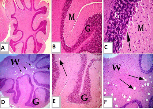
| Figure 5: (a-c): Section of mature Westar rats cerebellum of control group (a): showing normal, intact cerebellar cortex and medulla. (b): showing intact molecular layer (M) and granular layer (G). (c): showing normal molecular layer (M) and granular layer (G) and purkinje layer (arrow). (a-c) H&E a) 4X:10X. b) 10X:10X. c) 4X0: 10X. (d-f): Section of mature Westar rats cerebellum of exposed group to tartrazine (II). (d): showing numerous vacuolations of variable sized in cerebellar medulla (arrow), white matter (W) and gray matter (G). (e): showing slightly vacuolation in gray matter; cortex (arrow). (f): showing numerous vacuolations of variable sized in cerebellar medulla; white matter. d-f) H&E d) 4X:10X. e,f) 10X 10X. |