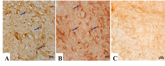
Figure 3:
Representative micrograph of immunopositive cells (arrows) in rat dental pulp. IGF-I (A), IGF-IR (B) and negative control (C). (Calibration bar: 5 μm).