 |
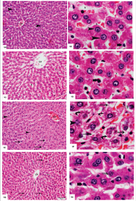
| Figure 1: Photomicrographs of sections in rats' livers stained with H and E. A. Group I (control group) shows a central vein (CV) with radiating cords of hepatocytes; most of them are mononucleated some are binucleated (arrowheads).Cords of hepatocytes enclose blood sinusoids (S) in between 200X. B. A higher magnification showing hepatocytes with acidophilic cytoplasm and central rounded vesicular nuclei (arrowheads). Blood sinusoids (S) are seen in between the cords of hepatocytes. Von Kupffer cells with dark nuclei are demonstrated (Thick arrow) 1000X. C. Group II (green tea group) demonstrates central vein (CV) with radiating hepatocytes. Note the blood sinusoids (S) 200X. D. A higher magnification showing the hepatocytes with blood sinusoids (S) in between. Note Von Kupffer cell (Thick arrow) 1000X. E. Group III (Ketamine group) reveals congested central vein (CV). Hepatocytes show cytoplasmic vacuolization (arrows). Some hepatocytes demonstrate dark nuclei (arrowheads) 200X. F. A higher magnification of group III showing hepatocytes with cytoplasmic vacuolization (arrows). Note the karryorhexed (arrowhead) and the small nuclei (double arrows). Deeply acidophilic fragmented cytoplasmic bodies are also noted (curved arrows) 1000X. G. Group IV (Ketamine and green tea group) shows central vein (CV) with radiating hepatocytes enclosing blood sinusoids (S) in between. Note some hepatocytes with deep acidophilic cytoplasm and dark small nuclei (arrows) 200X. H. A closer observation to the hepatocytes showing minimal cytoplasmic areas of vacuolization (arrow) and other with small nucleus (double arrow). No acidophilic fragmented bodies could be detected. Von Kupffer cell could be noted (Thick arrow) 1000X. |