|
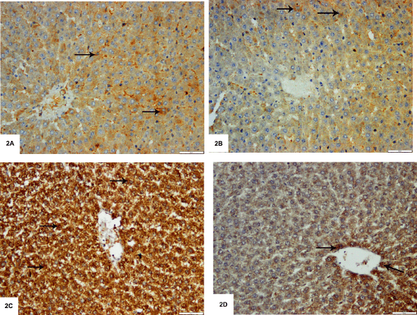
| Figure 2: Photomicrographs of sections of rats' livers stained with anti- iNOS antibody, 200X. A. The control group shows positive iNOS immunoreactivity within the cytoplasm of some hepatocytes (arrows). B. Group II demonstrates positive iNOS immunoreaction within the cytoplasm of some hepatocytes (arrows). C. Group III reveals positive iNOS immunoreactivity within the cytoplasm of nearly all hepatocytes (arrow). D. Group IV shows positive iNOS immunoreactivity within the cytoplasm of some hepatocytes (arrows). |