|
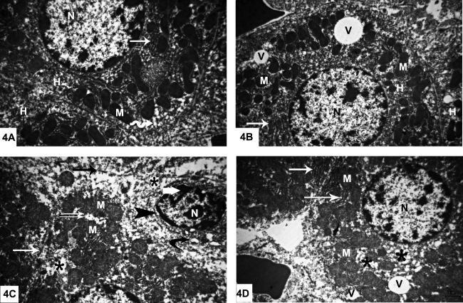
| Figure 4: Electron photomicrographs of rats’ livers, TEM, X8000. A. The control group shows two adjacent hepatocytes (H). One cell exhibits euchromatic nucleus with regular outline (N), many mitochondria (M), rough endoplasmic reticulum (arrow) and smooth endoplasmic reticulum (zigzag arrow). B. Group II demonstrates two neighboring hepatocytes (H). The hepatocyte has euchromatic nucleus (N), many mitochondria (M) and rough endoplasmic reticulum (arrow). Note two electron lucent vacuoles of variable sizes (V). C. Group III shows hepatocyte with shrunken nucleus with irregular outline (N), condensed peripheral heterochromatin (thick arrow) and focal areas of widened inter membranous space of nuclear envelope (arrowhead). Note multiple areas of cytoplasmic vacuolization (stars) around the nucleus and in relation to mitochondria. Cisternae of rough endoplasmic reticulum appear exhibiting discontinuity (arrow) or dilated (curved arrow). Mitochondria (M) are separated by empty spaces (double arrow) or discontinued rER (white arrow). D. Group IV shows hepatocyte exhibiting euchromatic nucleus with regular outline (N), many mitochondria (M) separated by regular (arrow) or widened rER (stars). Note two electron lucent vacuoles (V) of variable sizes with the smaller one near the cell surface. |