 |
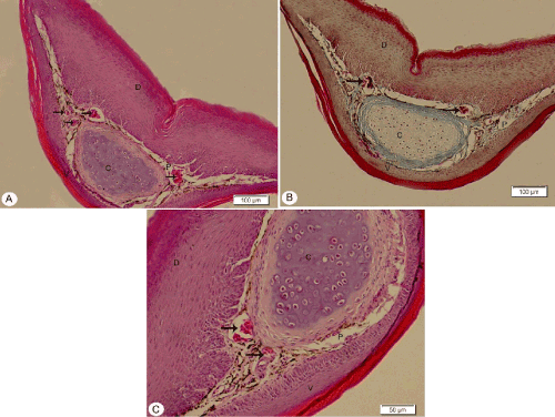
| Figure 3: Photomicrograph of a transverse section of the tongue apex showing the slightly keratinized dorsal surface (D), lamina propria (P), skeletal muscle fibers (arrows), highly keratinized ventral surface (V), cartilaginous plate (C) ,surrounded by large amount of collagenous fibers. A; H&E stain, B; Masson’s trichrome, C; high magnification of A. |