 |
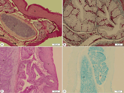
| Figure 4: Photomicrograph of transverse and longitudinal sections of the tongue body showing slightly keratinized dorsal surface (D), lamina propria (P), Faint PAS and strongly alcianophilic positive reactive tubuloaveolar lingual glands (G), skeletal muscle fibers (arrows), slightly keratinized ventral surface (V), Alcianophilic cartilaginous plate (C). A; H&E stain, B; Masson’s trichrome, C; PAS, D; Alcian blue. |