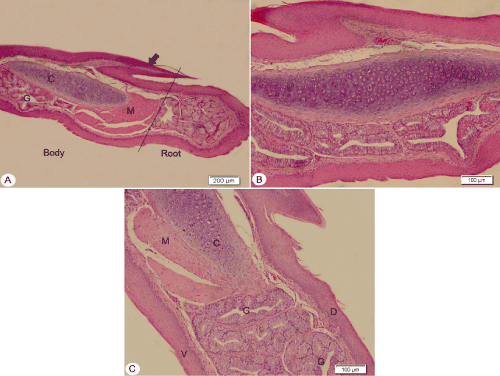
tongue body and root showing highly keratinized horny papilla (arrow),
lingual glands (G), skeletal muscle fibers (M), non-keratinized dorsal (D) and
ventral surface (V), cartilaginous plate (C).
 |
| Figure 5: Photomicrograph of longitudinal H&E stained sections of the tongue body and root showing highly keratinized horny papilla (arrow), lingual glands (G), skeletal muscle fibers (M), non-keratinized dorsal (D) and ventral surface (V), cartilaginous plate (C). |