 |
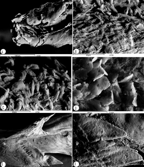
| Figure 6: Scanning electron micrograph of the dorsal surface of the tongue: A- apex, B, C and D- the rostral middle and caudal third of lingual body, E, Fbody and root, disquamated cells (d) leave-like at the apex and caudal third of tongue body while it is tap-like at the rostral and middle third of the tongue body, The central longitudinal groove (g), V- shaped row conical papillae between the body and the root of tongue (p1), additional row composed only of two larger papillae in each half behind the main row of papillae (p2), Root of tongue (r), corpus lingue (b) and Openings of the lingual slivary glands (o), larger peripherally located conical papillae located in the v- shaped row between the root and body of the tongue (p). |