|
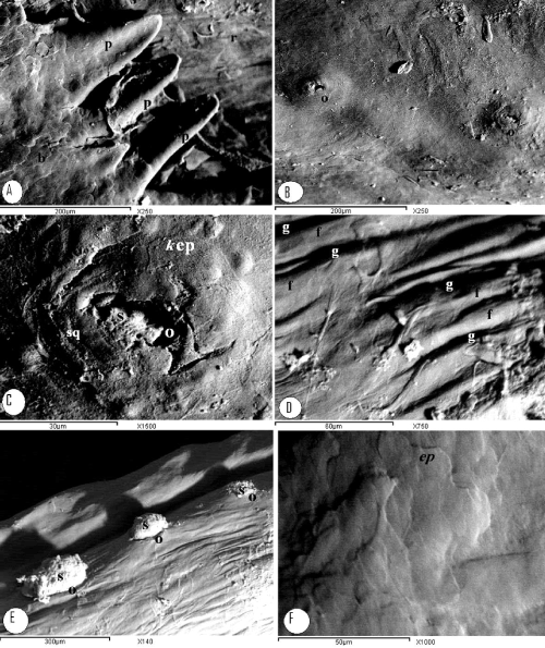
| Figure 7: Scanning electron micrograph of the dorsal (A-C) and ventral (D-F) surface of the tongue: A- body and root, B, C - root of the tongue, D, E- ventral surface of the body, F - tongue root, smaller medially located conical papillae (p) in the v- shaped row (b), elongated openings of the lingual salivary glands (o), Keratinized epithelium (kep), secretion (s), concentrically arranged epithelial cells (sq), longitudinal microridges (f) separated by longitudinal microgrooves (g), smooth non-keratinized epithelial surface (ep). |