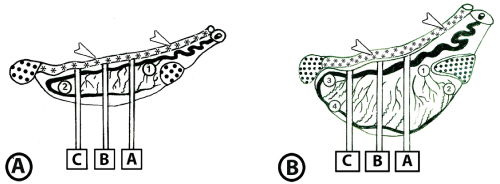
B: Diagrammatic representation of tissue sampling from left testis of premature and mature animals. Nineteen tissue blocks are collected from different areas of the head extremity (1, 2), tail extremity (3, 4) and three transverse sections (A, B, C). Slice B is taken from the testicular equator; sections A and C are equidistant from the equator and cranial or caudal pole, respectively. From each of the three slices five tissue blocks are obtained from the center, medial, lateral, epididymal and free sides.
• = Epididymis,*, arrowhead = Ductus deferens