 |
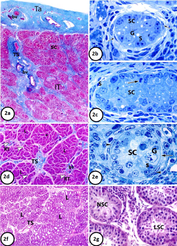
| Figure 2: 2A: Paraffin section in the of testis of newly born donkey showing tunica albuginea (Ta), testicular septum (Ts), semniferous cords (SC), interstitial tissue (IT) and blood vessels(bv). (Crossmon’s Trichrome, 25X). 2B: Semithin section in the testis of newly born donkey showing one seminiferous cord (SC) surrounded by one discontinuous layer of flattened cells (arrows), gonocytes (G) supporting cells (S). Notice the presence of variable- sized vacuoles within the germ and supporting cells. (Toluidine blue, 1000X). 2C: Semithin section in the testis of newly born donkey showing one seminiferouscord (SC). Notice, mitosis in gonocytes (arrow) (Toluidine blue, 1000X). 2D: Paraffin section in testis of suckling donkey (2 months old) was showing numerous irregularly distributed testicular lobules (L). Each lobule consists of tortuous seminiferous cords (*) and narrow interstitial tissue (arrow heads) in-between. Notice that the testicular septa (TS) are thick in the areas hosting testicular vessels (bv) and rete testis tubule (RT, arrow head). (Crossmon’s Trichrome, 50X). 2E: Semithin section in the testis of suckling donkey (6 months old) showing one semniferous cord (SC), gonocytes (G), supporting cells with indented nuclei (S) and peritubular cells (arrow head). Notice most of germ cells appear contacting the basement membrane (arrow). (Toluidineblue 1000X). 2F: Paraffin section in premature donkey testis (1.5 year) showing large number of irregular lobules (L) separated by irregular testicular septa (TS). The semniferous cords have increased both in length and convolutions. Notice some semniferous tubules show the primary signs of lumination are present besides the non-luminated and luminated ones. (Haematoxylin and Eosin, 50X). 2G: Paraffin section in premature donkey testis (1.5 year) showing non luminated semniferous cords (NSC) and luminated seminiferous cords (LSC). (HaematoxylinandEosin 400X). |