|
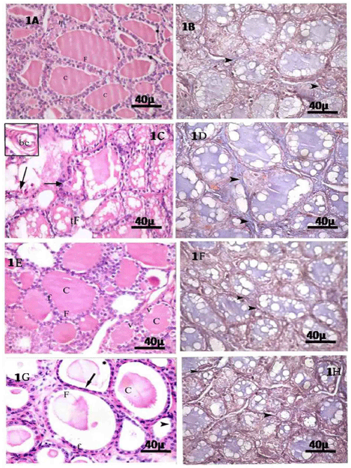
| Figure 1: control group (I); 1A, B: 1A: H&E stained section showing variable sized follicles. Central follicles (F) are lined by single layer of cuboidal cells with rounded nuclei (f) while the peripheral follicles have flattened or low cuboidal cells (*). Their lumina are filled with homogenous acidophilic structurless colloid (C) .It shows conspicuous peripheral vacuoles (v). Group of interfollicular cells are also observed (If). 1B mallory's trichrom stained section showing little collagen fibers (arrow head) in the connective tissue septa. EMF-exposed group (II) 1C, D: 1C: H&E stained sections showing disintegration and disorganisation of thyroid follicles with interrupted follicular wall (tF). Inset: congested blood vessels (bc) in C.T septa. 1D: mallory's trichrome stain section showing many collagen fibers (arrow head) in connective tissue septa. vitamin E-treated group(III) 1E,F: 1E: H&E stained sections reveal many recovered thyroid follicles (F) .These follicles are lined by cuboidal cells (f) with rounded nuclei. Note, return of peripheral vacuolation (v) in the colloid(C).1F: mallory's trichrome stained sections showing minimal collagen fibers (arrow head) in the connective tissue between the follicles. Recovery group (IV) 1G,H: 1G: H&E stained sections showing apparently normal variable sizes follicles (F). Some follicles appear distended (*) with colloid (C) and lined by flat cells (arrow). Other follicles have cuboidal cells (f) with rounded nuclei. Notice, there are involuted follicle (arrow head). 1H: mallory's trichrome stained sections showing moderate amount of collagen fibers (arrow head) in connective tissue septa between the follicles. (scale bar=40 µm ) |