 |
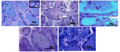
| Figure 2: Control group (I) 2A: Toluidine blue stained section reveal follicles (F) lined by cuboidal cells with rounded nuclei (f). These follicles are filled with colloid (C). Parafollicular C cell (pa) resting on the basement membrane have large pale nuclei (n).Inset: note the vascular connective tissue septa (vs) between the follicles. EMF-exposed group (II) 2B: Toluidine blue stained section showing disorganized thyroid follicles (df) with wide interfollicular spaces (S). Some follicular cells have dakely stained nuclei (n). Note, interfollicular cellular infilteration (If). 2C: demonstrate desquamated epithelial follicular cell (D). Vitamin E-treated group (III) 2D: Toluidine blue stained section reveal nearly normal thyroid follicles (F). They are filled with homogenous colloid (C) and lined by cuboidal cells (f) with vesicular rounded nuclei (n). Recovery group (IV) 2E: Toluidine blue stained section reveal variable shaped thyroid follicles (F), Some of them are involuted (arrow). Some follicles have darkely stained nuclei (*), others have normal cuboidal cells (f). Notice, wide intefollicular spaces (S) containing many blood vessels (bc) and cellular infilteration (If) (scale bar=20 µm) |