|
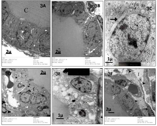
| Figure 3: Ultrathin sections of control group(I) 3 A,B,C :3A: showing part of follicle lined by cuboidal cells (f) that have rounded euchromatic nuclei (N (with clumps of heterochromatin (arrowhead).Their apical border shows microvilli (mv) projecting into the colloid (C). 3B: follicular cell cytoplasm shows dilated cisternae of rough endoplasmic reticulum (Rr), a few vacuoles (v) and also dense lysosomal granules (L).Note: blood capillary (bc) beneath the thin basal lamina (Bl) (Scale bar=2µm). 3C: showing parafollicular (pa) with a rounded euchromatic nucleus (N) lying on the basal lamina (arrow). Its cytoplasm contains many small electron-dense secretory granules (g), mitochondria (m), and also a few tubular cisternae of endoplasmic reticulum (Rr) (Scale bar=1µm). 3D, E, F: EMF-exposed group (II) 3D: showing high cuboidal follicular cells (f). These cells have rounded nuclei (N) with clumping of their peripheral heterochromatin. Dense lysosomal granule (L) and numerous vacuoles (V) are also seen. Their apical borders show microvilli (mv). Other follicular cells have electron dense shrunken nuclei (n). Dilated congested blood capillary (bc) and collagen fibers in interfollicular space (Scale bar=2µm). 3E: showing Numerous dilated irregular cisternae of rough endoplasmic reticulum (Rr) with loss of their lamellar arrangement(Scale bar=1µm).3F: demonsterate part of thyroid follicles (F) and interstitium (I)containing congested blood capillary (bc).Notice large amount of collagen fibers (arrow head) and fibroblast (arrow) (Scale bar=2 µm). |