 |
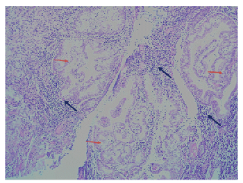
| Figure 1: A photomicrograph of a haematoxylin and eosin-stained section through the endocervix of the 88-year old woman treated with tamoxifen, showing typical histopathologic features of florid endocervical microglandular hyperplasia. Note the florid tightly packed endocervical glands lined by cuboidal cells associated with squamous metaplasia (red arrows). The stroma also shows infiltration by acute and chronic inflammatory cells close to the glands (blues arrows). Image was taken at 100x magnification. |