|
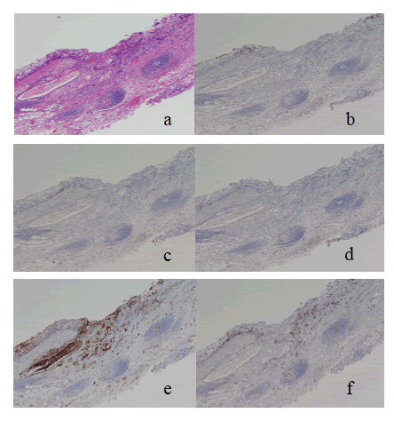
| Figure 4: Histology and immunohistochemistry of gastric cancer with C-pattern submucosal invasion. A case of moderately differentiated adenocarcinoma of type 0-IIc, measuring 12 mm in diameter, with submucosal invasion of >500 μm is shown. Both mucosal and submucosal layers are I-type. (a) HE staining (×100) (b) MUC5AC-negative (×100) (c) MUC6-negative (×100) (d) MUC2-negative (×100 (e) CD10-positive both in the mucosal and submucosal layers (×100) (f) MI Ki-67 is 61%. |