|
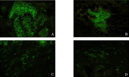
| Figure 2: (A) Negative-Localization of E-cadherin is along plasma membrane. (B) AGC: E-cadherin appeared to be diffusely distributed along the cell-cell contacts. (C,D). AC: E-cadherin was diffused into cytoplasm and E-cadherin expression was decreased. (Representative images of immunostaining for E-cadherin (green); ×200) |