|
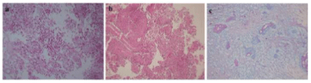
| Figure 2: a. Photomicrograph showing a glial tumor with prominent perivascular pseudorosettes (HE, X4) b. few halinized blood vessels with myxoid change in the background (HE, X10). c. Myxoid change highlighted by Alcian blue staining (Alcian blue, X20). |