 |
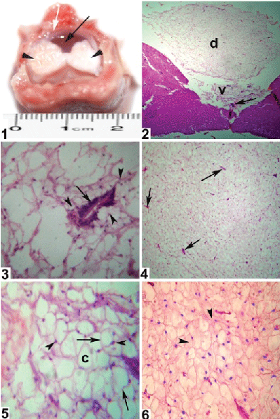
| Figure A1: Gross appearance of spinal cord L4 level glycogen body; delicate, ovoid circumventricular, transparent and gelatinous mass (black arrow), embedded in the dorsal part of the spinal cord lumbosacral region (arrow heads) and located inside the rhomboid sinus (white arrow). Figure A2: Glycogen body divided into dorsal (d) and ventral parts (v) and its ventral part appear enclosed the central anal (arrow) (H & E.X4). Figure A3: Spinal cord L4 level glycogen body showing the central canal (arrow) that completely enclosed with the glycogen body cells (arrowheads) (H & E.X40). Figure A4: Spinal cord L3 level glycogen body appeared with high vascularisation (arrows) (H & E.X 10). Figure A5: Spinal cord L3 level glycogen body polygonal cells (arrows), empty cytoplasm (C) and pushed nuclei toward one edge of the cells (arrowheads) (H & E X.40). Figure A6: Spinal cord L2 level glycogen body cells appeared filled with PAS +ve material (arrowheads) (PAS stain X.20). |