 |
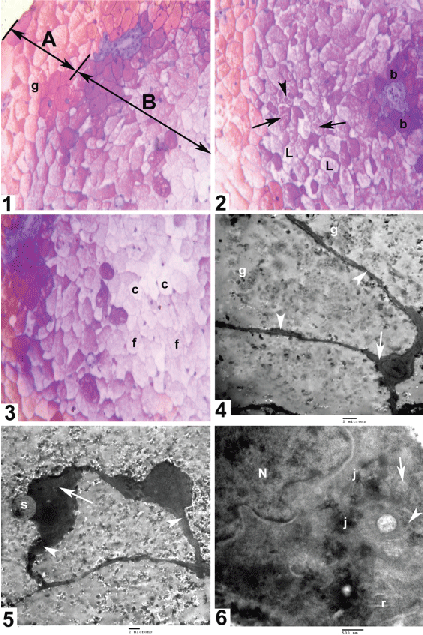
| Figure B1: Spinal cord L4 level glycogen body plastic section revealed peripheral faint red metachromatic cell zone (A) filled with granular eosinophilic material (g), and central basophilic cell zone (B) (Toluidine blue X40). Figure B2: Spinal cord L3 level glycogen body plastic section showing the outer part of the central zone with deep basophilic perivascular cells (b) and less basophilic outer ones (L). The cells are variable in size, most of them contain granular material (arrows) and few contain circular basophilic bodies (arrowhead) (Toluidine blue X40). Figure B3: Spinal cord L4 level glycogen body plastic section central zone most inner cells that had very light basophilic cytoplasm (f) and some appeared evacuated with intact cell membrane and peripheral nuclei (c) (Toluidine blue X40). Figure B4: Transmission electron micrograph showing the peripheral zone cells with small amount of juxtanuclear cytoplasm (arrow) and very thin cytoplasmic rim around the cell perimeter (arrowheads) and most of the cell filled with glycogen particles (g) (E/M X5000). Figure B5: Transmission electron micrograph showing the central zone cells with voluminous juxtanuclear cytoplasm (arrow) and broader cytoplasmic rim around the cell perimeter (arrowheads) and perinuclear circular membranous structure (s) (E/M X4000). Figure B6: Transmission electron micrograph showing the peripheral zone cells with electron dense nucleus (N), numerous polysomes clusters (arrowhead), mitochondria (arrow) rough endoplasmic reticulum (r) and cell junctions (j) (E/M X3000). |