 |
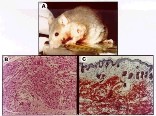
| Figure 1: Representative photographs of skins lesions in cholesterolfed apo E-KO mice. Panel A shows eruptive skin lesion on the shoulder area; Panels B and C present photomicrographs of the nuchal skin illustrating marked infiltration by lipids and presence of cholesterol granulomas and cholesterol clefting in Panel B (hematoxylin and eosin stain X50), and prominence of oil red O staining in Panel C (X25) [9]. |