|
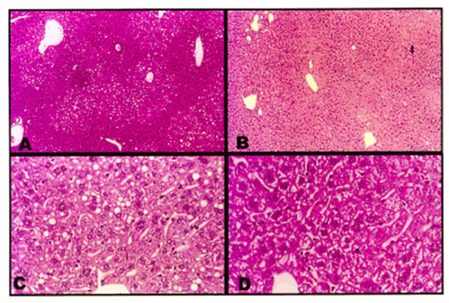
| Figure 3: Representative photomicrographs of livers from cholesterol-fed (Panels A and C) and one cholesterol+phytosterolfed (Panels B and D) apo E-KO mice. As is evident, non-specific vacuolation is predominantly seen in Panels A and D. Hematoxylin and eosin stain, A and B, X16; C and D X40 [9].. |