|
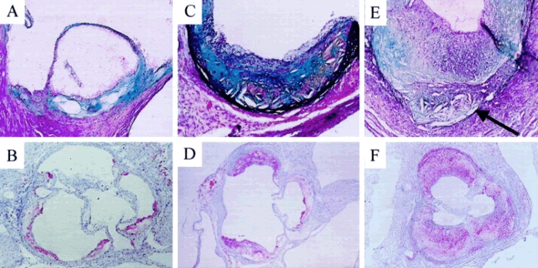
| Figure 4: Representative photomicrographs of serial transverse sections at the level of the aortic valve cusps taken from individual mice from FCP-3PI-treated (A and B), control (C and D), and probucol-treated (E and F) groups. The volume and complexity of the atherosclerotic lesions are markedly reduced in A and B and increased in E and F, respectively, compared with C and D. Whereas the ORO stain (B, D, and F) emphasizes lipid accumulation in the lesions, Movat pentachrome stain (A, C, and E) highlights components of the lesions including interstitial matrix, foam cells, and cholesterol clefts. E also illustrates an area of aortic wall destruction with evidence of apparent early aortic aneurysm formation (arrow) (original magnification ×25, B, D, and F; A, C, and E ×50). FCP-3PI was a phytosterol mixture [14]. |