|
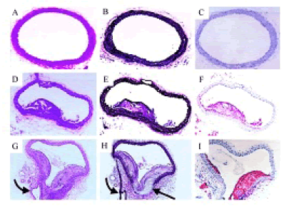
| Figure 5: Representative photomicrographs of transverse sections of thoracic aortas from individual mice from FCP-3PI-treated (A, B, and C), control (D, E, and F), and probucol-treated (G, H, and I) groups stained with hematoxylin and eosin (A, D, and G), Movat pentachrome (B, E, and H), and ORO (C, F, and I). Sections from FCP-3PI–treated animals show no visible intimal lesions and normal musculoelastic layers. Sections from both control and probucol-treated mice reveal advanced atherosclerotic lesions containing foam cells, cholesterol clefts, and increased interstitial matrix. As is apparent, the nature of the lesions is more complex in the probucol-treated animal. H shows complete disruption of media (straight arrow) at a bifurcation point (curved arrow) in the probucol-treated mouse (A through F, original magnification ×25; G through I × 50). FCP-3PI was a phytosterol mixture [14]. |