 |
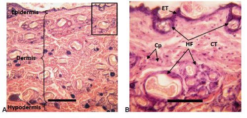
| Figure 6: A) Normal skin rabbit stained with Hematoxylin-eosin. Slice is perpendicular to the skin surface (on top). Scale bar: 300 μm. B) Magnification of the square area in A. Scale bar: 100 μm. Epidermis: Epithelium tissue (ET) of cells stained in purple (hematoxylin). Hair follicles (HF) are on surface. Dermis: Loose connective tissue (CT) with isolated fibroblast nuclei in purple, collagen fibres and elastic fibres cut in rose in different spatial directions and ground substance in white between abundant hair follicles (HF: white spaces with several pink eosin hairs surrounded by purple epithelium) and some capillaries (Cp). No evidence of inflammatory reaction (abundant very purple leukocytes) neither fibrosis (dense connective tissue with a predominance of very pink collagen fibres). Hypodermis: Connective tissue under the dermis. |