|
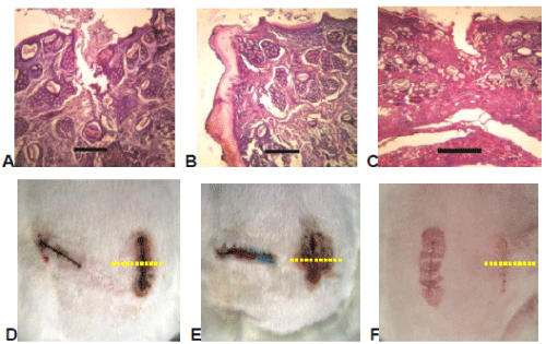
| Figure 7: Histopathological and clinical appearance of wounds closed with ECN (A,D), BCN (B,E) and OCN (C,F) adhesives after 3 days. Right side of rabbit is on the right of pictures (D,E,F). Yellow dotted lines: cross sections of wounds corresponding to slices with skin surface on the top. Scale bar: 300 μm. The application of the ECN and OCN produced wound edges lightly separated on the surface but joined at the bottom (Degree 3 in Table 1) (A and C pictures). In B picture one edge of open wound with remains of adhesive is shown (Degree 1 in Table 1). |