|
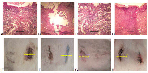
| Figure 8: Histopathological and clinical appearance of wounds closed with ECN (A,E), BCN (B,F) and OCN (C,G) adhesives and suture (D,H) after 7 days. Right side of rabbit is on the right (E,F,G,H). Yellow dotted lines: cross sections of wounds corresponding to slices with skin surface on the top. Scale bar: 300 μm. Pictures A, C and D show that the use of ECN or OCN adhesive renders the wound closed and the skin tissue shows almost normal structure (degree 5 in Table 1) as compared to the use of suture (Degree 4 in Table 1). When BCN adhesive is used (B picture), the wound edges are totally separated (Degree 2 in Table 1). |