|
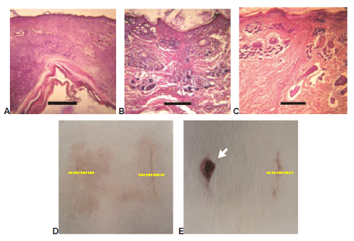
| Figure 9: Histopathological and clinical appearance of wounds closed with BCN (A,D-left), OCN (B,D-right) and adhesives and suture (C,E) after 14 days. Right side of rabbit is on the right (D,E). Yellow dotted lines: cross sections of wounds corresponding to slices with skin surface on the top. Arrow: open wound. Scale bar: 300 μm. In B picture the skin shows almost normal structure with very little evidence of fibrosis (Degree 6 in Table 1) as compared with A (Degree 4 in Table 1) and C (Degree 5 in Table 1) pictures. |