 |
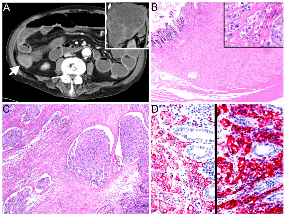
| Figure 1: Clinicopathologic features of hepatic epithelioid hemangioendothelioma with cecal metastasis.(A) Abdominal CT reveals an ileocecal mass (arrow), measuring 4.0x3.3 cm with small bowel obstruction. Extensive tumor involvement in the liver is noted (inset). (B) Microscopically, the tumor infiltrates the colonic wall with a focal mucosal involvement (H&E, 20X), and is composed of cords or nests of epithelioid cells with characteristic intracytoplasmic vacuoles in a myxohyaline stroma (inset, H&E, 400X). (C) Extensive tumor emboli are found in the sample (H&E, 100X). (D) Immunohistochemically, the tumor cells are positive for CD34 (left panel) and CD31 (right panel). |