|
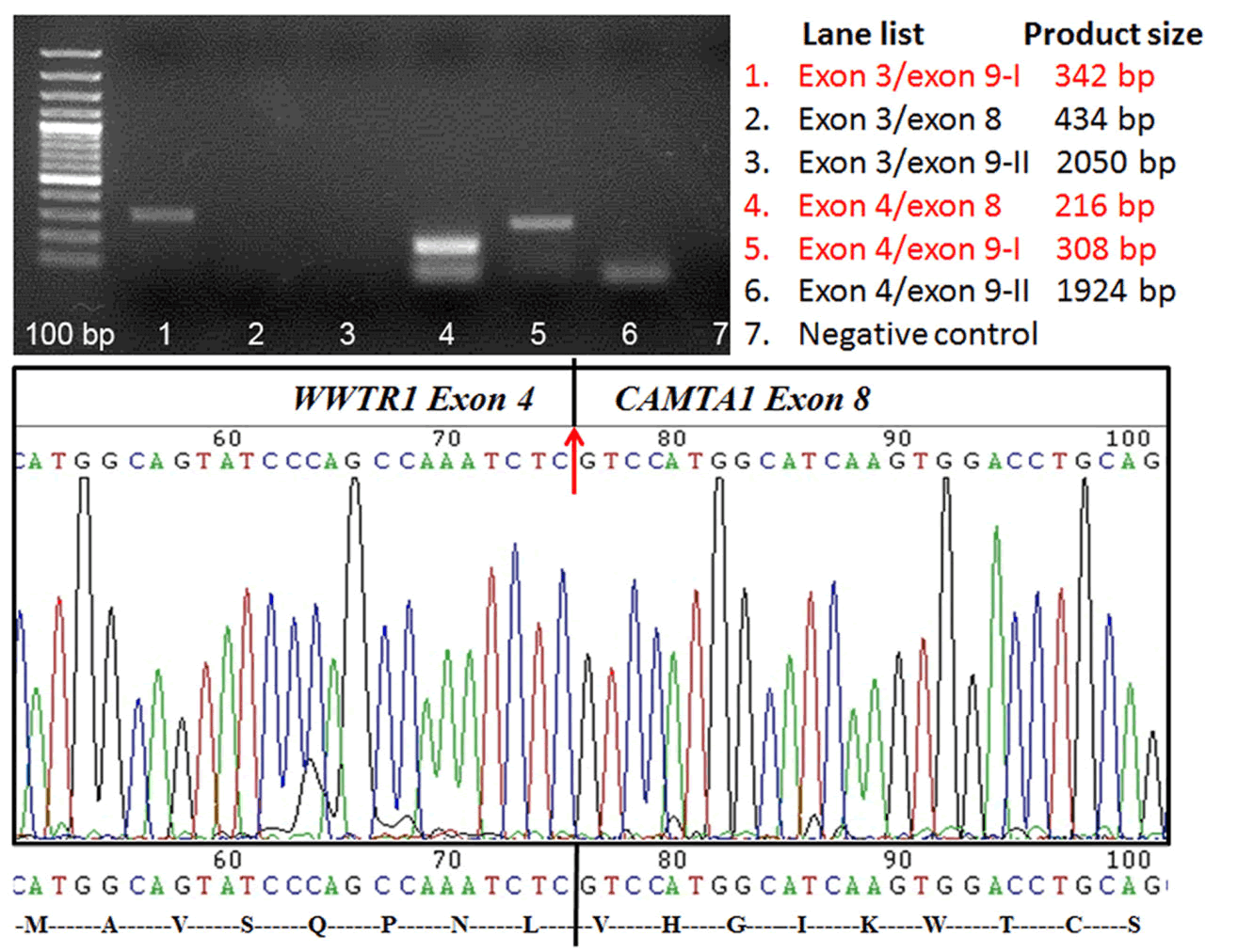
| Figure 2: Molecular studies of the WWTR1-CAMTA1 gene fusion. Paraffin-embedded tumor tissues were used for RT-PCR to test for the WWTR1-CAMTA1 fusion transcript with six pairs of primers as listed. The positive results are labeled in red (upper right panel). Because the RNA molecules were fragmented in paraffin tissue, expected products more than 400 bp were unamenable for detection (upper left panel lanes 2, 3 and 6). The lower bands in lanes 4 and 6 indicate non-specific PCR products. The fusion involves exon 4 of WWTR1 with exon 8 of CAMTA1 as detected by Sanger sequencing (lower panel). |