|
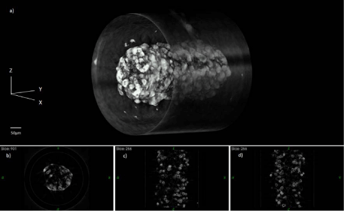
| Figure 4: Volume rendering (a) and slices (b, c, d) of one field of view of Muntjac cells stained with hematoxylin inside a 320 μm ID capillary tube as seen under 10x objective, rendered using Volview. Reconstruction can be rotated to be seen in any orientation. Slices can be obtained along the xy (b), xz(c), and yz(d) planes. |