|
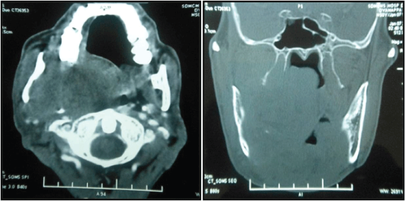
| Figure 3: A) Axial CT with contrast showing a well-defined heterogeneously enhanced soft tissue mass involving the masticator and parapharyngeal spaces, causing anterior and lateral displacement of ramus and angle of the mandible on right side and predominantly compromising the oro-pharyngeal airway column. B) Bone window section of CT showing cortical thinning and erosion of the lingual surface of ramus and angle of the mandible on the right side. Medially, the mass is displacing the lateral pharyngeal wall and involving the pterygopalatine fossa with significant narrowing of naso and oro-pharyngeal airway column. |