|
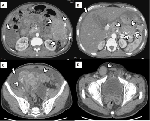
| Figure 1: Axial MDCT scan images of the abdomen acquired in the arterial (A,B) and venous phases (C,D) demonstrating multiple bulky, lobulated, heterogeneously enhancing, intraperitoneal soft tissue masses in the left para-colic gutter, lesser sac and pelvis (arrowheads in A, B and C, respectively) with moderate ascites (arrows). Similar soft-tissue lesions were seen diffusely scattered in the entire peritoneal cavity (not shown). Associated nodular thickening of the parietal peritoneum was present which was extending into the right inguinal canal (arrowhead in D). |