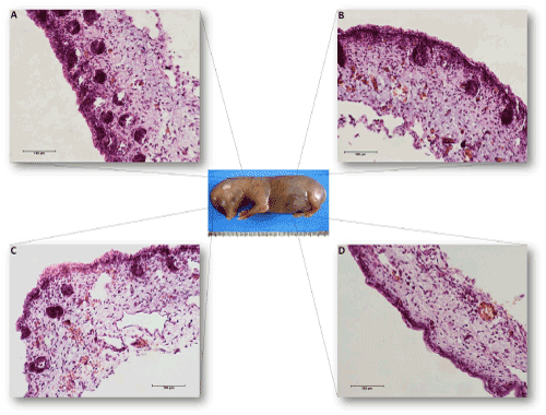
Figure 3:
An illustrative scheme of the skin micrographs in different body regions of the 44-day-old canine fetus. (A) the dorsal cervical region; (B) the dorsal lumbar region; (C) the head; and (D) the ventral abdominal region.