|
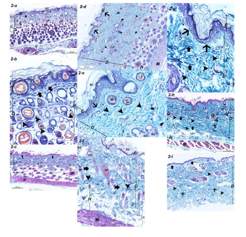
| Figure 2: Skin of the different age groups stained with Gomori’s trichrome stain showing the stratum corneum (SC) of the epidermis (E), dermis (D), hypodermis (H), hair follicles (HF), sebaceous glands (SG) and a layer of skeletal muscle (M) beneath the hypodermis. Note and fat sells (*). (a) the skin of a ten-day old rat. The collagen fibres in the papillary layer (arrow) and reticular layer (arrowheads) were sparse (Gomori’s trichrome stain 100X). (b) Higher magnification of the previous figure (Gomori’s trichrome stain 400X). (c) the skin of a twenty day old rat. The collagen fibres in the papillary layer (arrows) and reticular layer (arrowheads) distributed parallel to the epithelial surface thick and were regularly arranged (Gomori’s trichrome stain 100X). (d) the skin of a two month old rat. The collagen fibres in the papillary layer (long arrows) were parallel to the epithelial surface, while that of the reticular layer were arranged in the form of bundles. These bundles were distributed parallel (arrowheads) and perpendicular (short arrows) to the surface epithelial (Gomori’s trichrome stain 100X). (e) Higher magnification of the previous figure (Gomori’s trichrome stain 400X). (f) the skin of a six month old rat. The collagen fibres in the reticular layer were arranged in thick bundles (arrowheads) parallel to the surface epithelial and thin bundles (arrows) perpendicular to the surface epithelial. (Gomori’s trichrome stain 100X). (g) Higher magnification of the previous figure (Gomori’s trichrome stain 400X). (h) the skin of a one year old rat. The collagen fibres in the papillary layer (*) were thin and parallel to the epithelial surface. The collagen fibres in the reticular layer were arranged in thick bundles (thick arrows) parallel to the surface epithelial. Two forms of bundles perpendicular to the surface epithelial were found; thick bundles (thin arrows) and thin bundles (arrowheads). (Gomori’s trichrome stain 100X). (i) the skin a two year old rat. The collagen fibres in the papillary layer (thick arrows) were very thin and parallel to the epithelial surface. The collagen fibres in the reticular layer were arranged in thick bundles (thin arrows) parallel and thin bundles (arrowheads) perpendicular to the surface epithelial. Note atrophy of the hair follicles and absence of the layers of the stratum corneum (Gomori’s trichrome stain 100X). |